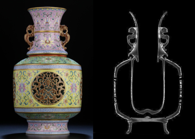
X-rays are a form of electromagnetic radiation, similar to visible light. The difference between visible light and x-rays is the wavelength. Unlike light, x-rays have higher energy with shorter wavelengths and can pass through most objects. Detectors on the other side of the object capture the x-rays, resulting in a two dimensional image. X-ray images will show forms superimposed, i.e. the structure in the front will be superimposed over those in the back, so the whole structure will be seen layered at one time.
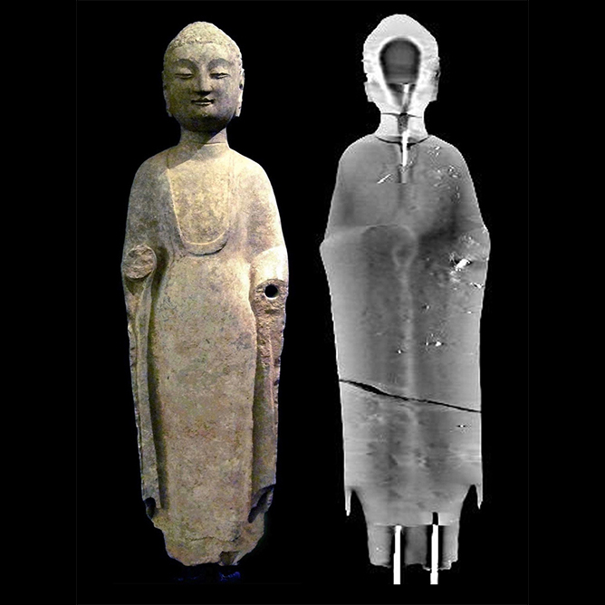
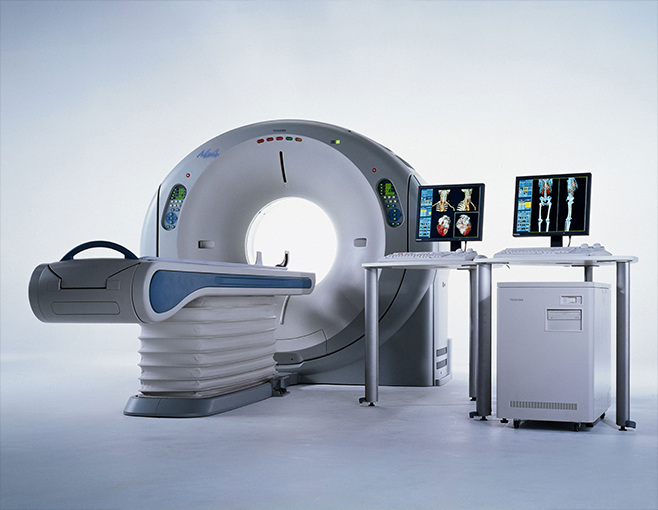
CT (computed tomography) is a medical imaging method using an X-ray source and detector unit rotating synchronously around an object. Data is acquired continuously during rotation and when reconstructed can provide cross-sectional and three dimensional views. CT scans can also show smaller contrast differences than conventional x-ray images, and this allows more detailed visualization of the inner state of a work of art, i.e. condition, areas of restoration or repair, method of construction, and natural damage such as oxidation, corrosion or cracking.
Although CT Scanning and X-ray are not in themselves dating methods, they do not affect levels of TL or 14C in an object and can be safely used in conjunction with other such techniques of observation and analysis to provide more complete information in the process of authentication.
Most kinds of materials can be CT scanned or x-rayed, although the size of the gantry opening on the CT machine limits the size of the objects that can be scanned.
X-rays use radiation to produce images of an object. Dense areas block the radiation which appear white in images and show forms superimposed, i.e. the structure in the front will be superimposed over those in the back so the whole structure will be shown layered at one time.
While traditional x-ray machines image structures in two dimensions, CT scans allow for a more three-dimensional effect. CT employs tomography (imaging by sections) created by computer processing and produces 360 degree views.
Digital geometry processing is used to generate three dimensional images of the inside of an object from a large series of two dimensional x-ray images taken around a single axis of rotation. A volume of data is produced that can be manipulated, through a process known as windowing, to show various structures based on their ability to block the x-ray beam. This volume of data can be reformatted in various planes or even as volumetric, or three dimensional, representations of the structures.
Due to the inherent high-contrast resolution of CT, differences between structures that differ in physical density by less than 1% can be distinguished. Referred to as multiplanar reformatted imaging, data from a single CT imaging procedure consisting of either multiple, contiguous or one helical scan can be viewed as images in the axial, coronal, or sagittal planes.
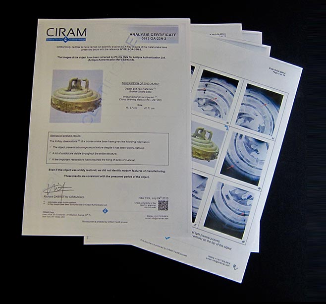
The scan or x-ray images are reviewed and reported on by a specialist radiologist with extensive training and background in sectional interpretation and x-ray imaging. A full report can be provided that includes relevant x-ray images or relevant axial, sagittal and coronal slices, 3D reconstructions, opaque and transparent views, as well as observations on construction, any areas of interest and conclusions.
 中文
中文 ENGLISH